



A very small incision. No stitches in most cases. And a visual recovery that many patients begin to notice from the very next day. Phacoemulsification is the technique that has transformed cataract surgery and allows the opacified lens to be removed with precision using ultrasound.
Modern surgery, however, goes beyond using an advanced machine. It integrates the size and architecture of the incision, the control of fluids inside the eye, the energy used, the planning of the intraocular lens and, in selected cases, the femtosecond laser. The goal is the same for every patient: to treat the cataract safely and to guide the procedure toward the best possible visual quality according to their needs.

What is phacoemulsification
Phacoemulsification is a surgical technique used to remove the lens when it has become opaque due to a cataract. The term describes what happens during the operation: a very thin probe emits ultrasound to break the lens into tiny particles and aspirate them through a microincision.
Unlike classic extracapsular extraction, which required a much larger wound to remove the lens in one piece, this technique preserves the capsular bag and allows a foldable intraocular lens to be implanted in it. The National Eye Institute notes that most current cataract surgeries are performed using phacoemulsification; it is also the procedure described by Mayo Clinic as the standard of modern cataract surgery.
Why the lens becomes opaque
The most common cause of cataract is natural aging. Poorly controlled diabetes, ocular trauma, certain prolonged treatments with corticosteroids and other individual factors can also accelerate its onset. The opacity may be located mainly in the center of the lens —nuclear cataract—, at its edges —cortical— or in its posterior zone —posterior subcapsular—. Each type can affect night driving, reading, contrast or glare differently.
Not every cataract must be operated on as soon as it is diagnosed. The decision is usually made when vision begins to limit activities that are relevant to the person, such as reading, driving, working or recognizing faces. In the article on when to operate on cataracts the criteria assessed in consultation are explained.
How the surgery is performed, step by step
The procedure is usually outpatient. Topical anesthesia with drops is commonly used and, when necessary, light sedation. It usually does not require hospital admission.
- Corneal microincision. The surgeon creates an entry at the edge of the cornea. In conventional phacoemulsification it usually measures approximately between 2 and 3 mm; with specific instruments and lenses, in selected cases microincisions smaller than 2 mm can be performed, even close to 1.1 mm.
- Capsulotomy or capsulorhexis. A circular opening is made in the capsule that surrounds the lens. Its size, regularity and centration are important for the stability and final position of the intraocular lens.
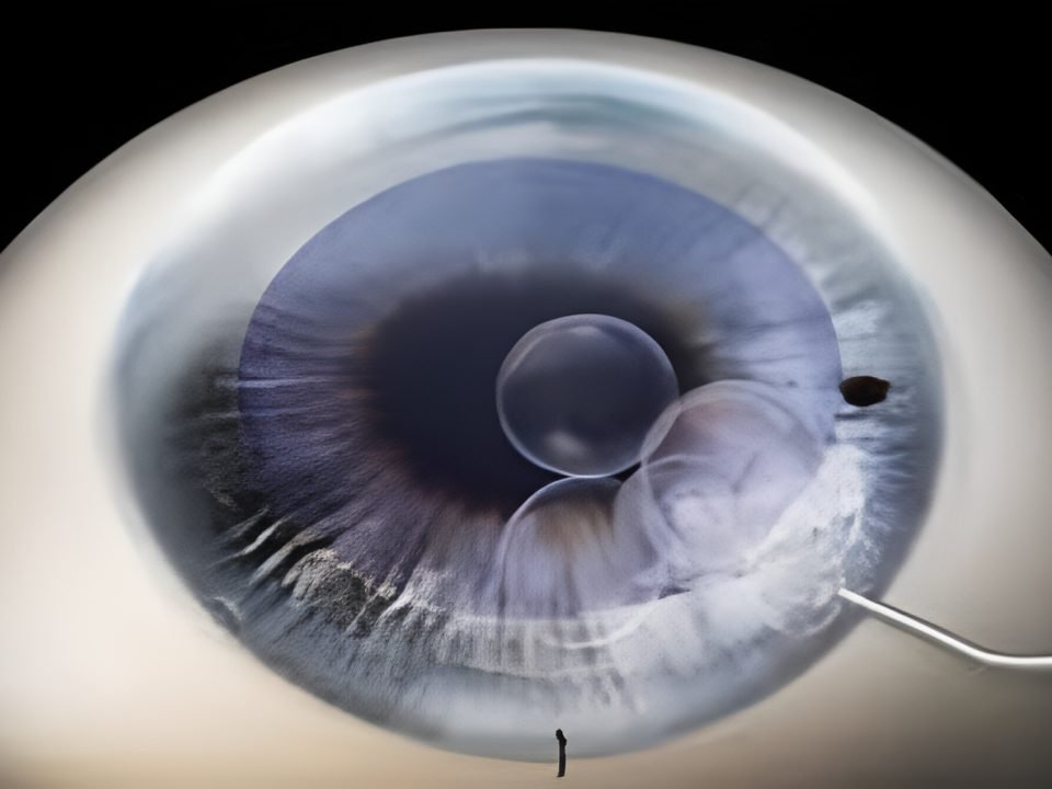
- Fragmentation and aspiration. The phacoemulsification probe fragments the lens using ultrasound and aspirates the material in a controlled manner, preserving the capsular bag.
- Intraocular lens implantation. Once the cataract has been removed, a foldable lens is inserted and unfolds inside the capsular bag.
- Self-sealing closure. The small incision usually seals on its own, so most procedures do not require stitches.
The surgical procedure usually lasts between 10 and 20 minutes per eye, although the total time at the center includes preparation, surgery and immediate recovery.
Microincision: precision that must be adapted to each eye
A small incision is not an isolated goal: it must be appropriate for the instruments, technique and lens chosen. When well planned, it can reduce alteration of corneal architecture, minimize induced astigmatism, promote self-sealing closure and contribute to rapid functional recovery.
The philosophy of high-precision surgery does not consist of always making the smallest possible incision, but rather choosing the safest and most appropriate size for each eye. Cataract density, the cornea, the anterior chamber and the type of lens are part of that decision.
Fluidics control and ultrasonic energy
During phacoemulsification, the surgeon controls irrigation, aspiration, intraocular pressure, anterior chamber depth and ultrasonic energy. Modern fluidics systems help maintain more stable conditions while the cataract is fragmented and aspirated. This allows the surgery to be adapted to the lens density and the anatomical characteristics of each patient.
Reducing, when possible, the time and energy of ultrasound is relevant because the lens is surrounded by delicate structures, including the corneal endothelium. It is a goal of tissue protection, not a promise of zero risk: the response of each eye also depends on the cataract, the cornea and the complexity of the procedure.
Phacoemulsification and femtosecond laser
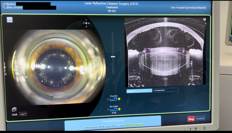
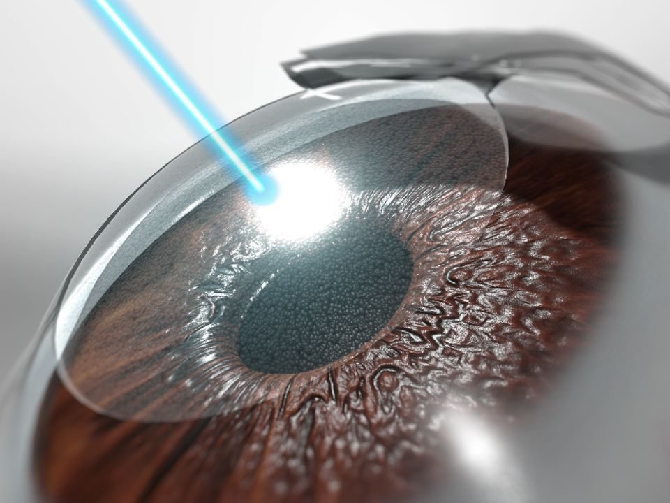
The femtosecond laser does not replace phacoemulsification: the extraction and aspiration of the material is usually completed using ultrasound. It can assist specific stages, such as the corneal incision, capsulotomy, initial fragmentation of the lens and, in certain cases, incisions for the treatment of astigmatism.
Its main value is the reproducibility of these stages and the possibility of reducing the phaco energy needed after pre-fragmenting the lens. Comparative evidence has observed reduced energy and effective phacoemulsification time, as well as greater precision in capsulotomy parameters. Both approaches—with laser or conventional phacoemulsification—are effective and safe; the practical difference depends on the specific indication, not on a technology applied indiscriminately.
You can learn more about this technology in the article on femtosecond laser in eye surgery.

Why intraocular lens centration matters
The intraocular lens must be well calculated, correctly positioned, stable, and centered. This is especially important when implanting toric, multifocal, or extended depth of focus (EDOF) lenses, because their optical outcome can be more sensitive to position and, in toric lenses, to the alignment axis.
A regular and centered capsulotomy contributes to that goal. In certain cases, digital assistance systems, such as CALLISTO eye, may be used to facilitate planning and alignment of toric lenses. Technology provides tools; lens selection and integration of all parameters remain individualized clinical decisions.
Intraocular lenses: a decision before surgery
The cataract extraction technique is similar, but planning changes according to the visual goal. Monofocal lenses offer maximum sharpness at one focus, usually distance, and often require glasses for near vision. Multifocal or EDOF lenses can reduce dependence on glasses, although some people perceive night halos during the adaptation process. Toric lenses correct corneal astigmatism and can be combined with other optical solutions.
The choice is based on refraction, corneal and retinal status, ocular surface, lifestyle, and visual expectations. The premium intraocular lenses require especially precise planning. In many cases, the ocular biometry calculates lens power and the corneal topography helps assess astigmatism and corneal regularity. You can consult an additional explanation about the different types of intraocular lenses and about the correction of presbyopia.
Recovery and care after phacoemulsification
Recovery is usually quick, but it is not identical for everyone. During the first 24 hours, there may be somewhat blurred vision, light sensitivity, or a mild foreign body sensation. Most patients notice early improvement; complete visual stabilization may take two to four weeks, depending on the eye, the lens, and individual progression.
- Use antibiotic and anti-inflammatory eye drops exactly as prescribed. If you use several, leave at least five minutes between them.
- Do not rub your eye or sleep with your face on the operated side during the first few days.
- Avoid pools, the beach, and intense physical exertion during the period indicated by the medical team.
- Wear sunglasses outdoors if you are sensitive to light.
Severe pain that does not subside, sudden vision loss, sudden flashes of light, or purulent discharge are not expected symptoms: they require contacting the clinic without waiting for the scheduled follow-up. If you are concerned about which discomforts are common, see the article Is eye surgery painful?.

What active fluidics provides
During phacoemulsification, aspiration of material and irrigation that maintains the anterior chamber change continuously. Active fluidics allows dynamic adjustment of irrigation, aspiration, and intraocular pressure to maintain a more stable anterior chamber. In some systems, this helps approximate intraocular pressure to the programmed value even when aspiration conditions change.
The purpose is to provide a more controlled surgical environment and adapt parameters to cataract density, ocular anatomy, and each phase of the procedure. It does not replace the surgeon's judgment: it is a tool that is part of a personalized phacoemulsification strategy.
Corneal endothelium protection
The corneal endothelium is a delicate cell layer that helps maintain corneal transparency. Ultrasonic energy, surgical time, cataract density, and prior corneal characteristics can influence the stress it receives during surgery.
Prior fragmentation with femtosecond laser can reduce energy and effective phacoemulsification time in selected procedures. Some studies have observed favorable differences in endothelial loss, but the result is not uniform across all studies or all patients. Therefore, it should be understood as a possible advantage of energy reduction, not as a guarantee of endothelial preservation.
Recovery: which factors truly influence it
Incision size and architecture are only part of recovery. Cataract density, ultrasonic energy used, corneal and ocular surface condition, retina, type of implanted lens, and individual patient characteristics also influence it.
The combination of microincision, optimized phacoemulsification, advanced fluidics, and femtosecond laser in selected cases aims to reduce surgical aggression factors and promote rapid functional recovery. Even so, the visual difference compared to conventional surgery is not necessarily large or persistent in all people.
A precision strategy, not a single device
Modern surgery integrates advanced biometry, topography, and, when indicated, corneal tomography and ocular surface analysis. This is combined with microincision, dynamic fluidics control, femtosecond-assisted capsulotomy and fragmentation, digital assistance for aligning certain lenses, and personalized selection of the intraocular lens.
In patients with toric, multifocal, or EDOF lenses, calculation, position, stability, centration, and, when applicable, lens orientation are relevant variables. CALLISTO eye is a digital assistance system that can help the surgeon identify and track the planned axis during toric lens implantation; it does not replace planning or surgical technique.
Microincision and surgical experience in Marbella
Dr. Ali Nowrouzi has incorporated phacoemulsification techniques with microincisions smaller than 2 mm at centers on the Costa del Sol and Campo de Gibraltar, when patient characteristics, instrumentation, and the selected lens allow it. The choice of incision size seeks to balance safety, anterior chamber stability, surgical ease, and refractive outcome.
Technology provides tools, but integrating microincision, fluidics, femtosecond laser, advanced biometry, premium lens, and precision centration requires experience in cataract surgery and refractive surgery.
Technology and experience: two inseparable elements
High-precision cataract surgery combines diagnosis, biometry, topography, microincision, fluidics, phacoemulsification, femtosecond laser in selected patients, and a custom-chosen intraocular lens. No device replaces clinical judgment or surgeon experience.
Dr. Ali Nowrouzi holds the FEBOS-CR certification (Fellow of the European Board of Ophthalmology – Cataract and Refractive Surgery), specific training in refractive and lens surgery, and experience at centers such as Quirónsalud Marbella, Hospital Ochoa, Quirónsalud Campo de Gibraltar, and Premium Clinic Marbella. You can learn about his career on the page about Dr. Ali Nowrouzi.
Frequently asked questions
Does phacoemulsification hurt? It is performed with topical anesthesia and, in most cases, no pain is felt during the procedure, although slight pressure may be noticed. Subsequent discomfort is usually mild.
Will I need stitches? Usually not. The microincision is typically self-sealing, although the decision depends on the surgical conditions of each case.
Is femtosecond laser necessary for all cataracts? No. It can provide value in selected cases, but its indication depends on the eye, the cataract, the visual goal, and the surgical strategy.
Does microincision guarantee faster recovery? It may favor less corneal alteration, but recovery depends on many factors and cannot be guaranteed solely by incision size.
Do I have to wait for the cataract to “mature”? No. A very advanced cataract may require more energy and make surgery more complex. In general, surgery is considered when symptoms already limit daily life.
What is active fluidics? It is a system that dynamically adjusts irrigation, aspiration, and intraocular pressure during phacoemulsification. Its function is to help maintain more stable conditions inside the eye while the cataract is removed.
What does CALLISTO eye provide? It is a digital assistance tool that can help track the planned implantation axis of a toric lens. It is useful within individualized planning, but it does not replace prior calculation or the surgeon's technique.
Request a personalized assessment
If you have been diagnosed with a cataract and want to know which technique and which lens may best suit your case, a complete ophthalmological assessment allows reviewing your visual history, performing the necessary diagnostic tests and establishing individualized planning. You can book an appointment here.
Scientific evidence and references
The choice between conventional phacoemulsification and femtosecond laser-assisted surgery must be individualized. Published evidence shows advantages of the laser in several surgical precision parameters, but it does not allow promising a superior visual outcome for all patients.
2024 meta-analysis (23 trials, 4,844 eyes). Pichardo-Loera and colleagues compared femtosecond laser-assisted cataract surgery (FLACS) with conventional phacoemulsification. The analysis found statistically significant differences in favor of FLACS in capsulotomy circularity, intraocular lens centration, cumulative dissipated energy, mean phacoemulsification time, and effective phacoemulsification time. The authors also reported a difference in endothelial density loss; the clinical significance must be interpreted in the context of each patient and the overall body of evidence. View study on PubMed.
2025 systematic review (41 trials, 9,310 eyes). Song and colleagues found a more precise capsulotomy, a more optimized effective lens position, and a lower incidence of intraocular lens decentration with FLACS. Both procedures were considered effective and safe. The analysis reported advantages in some early and long-term visual outcomes, while no visual difference was found at intermediate follow-up. View study on PubMed.
Clinical interpretation. The American Academy of Ophthalmology concluded in its technology assessment report that both techniques offer excellent safety and refractive outcomes, without an overall superiority of one over the other. Therefore, the laser should be considered as a tool that may be useful in selected cases —for example, depending on ocular anatomy, cataract type, or the planned lens—, not as a universal guarantee of better vision or recovery. View report on PubMed.