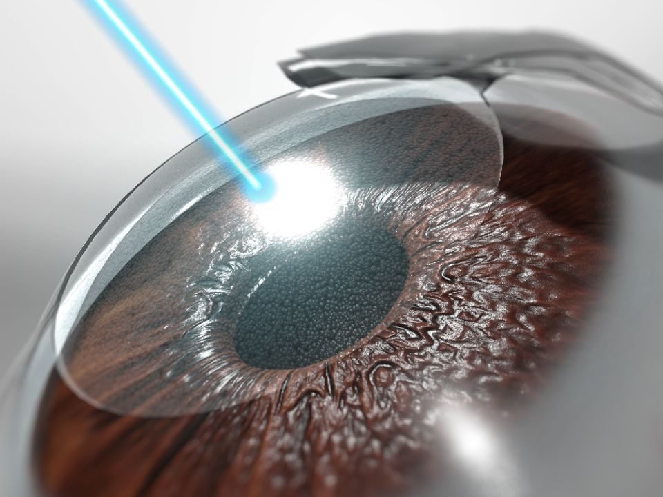

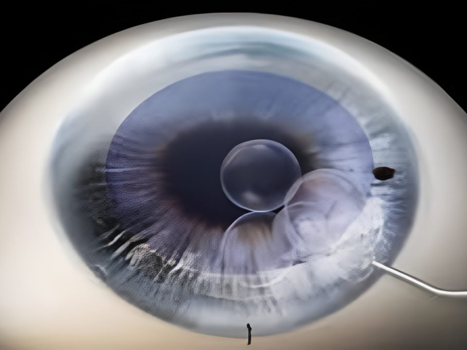

One in ten keratoconus patients has a parent who also suffered from it, and just two decades ago, most advanced cases ended in a corneal transplant. In Dr. Ali Nowrouzi's practice in Marbella, that reality has changed: today there is a technique that replaces synthetic rings with segments of donated human corneal tissue, implanted with the precision of a femtosecond laser.
It is called Femto-CAIRS (Femtosecond Laser-Assisted Corneal Allogenic Intrastromal Ring Segments) and it is marking a real change in how keratoconus moderate and advanced keratoconus is treated.
Keratoconus is a progressive corneal disease that usually appears in adolescence or young adulthood. The corneal tissue progressively thins and takes on a conical shape. Light no longer focuses correctly on the retina, causing blurred vision, irregular astigmatism, halos, and glare.
The Corneal Cross-Linking made it possible to halt disease progression in thousands of patients. Then came synthetic intracorneal segments, which improved corneal shape and reduced irregular astigmatism. Femto-CAIRS is the next step: it changes the implanted material to human corneal tissue, with more natural biological integration.
What is keratoconus and why does it compromise vision
Keratoconus is a non-inflammatory corneal ectasia. The cornea progressively loses its biomechanical resistance: as collagen fibers weaken, intraocular pressure causes it to take on a conical and irregular shape. This alters the way light passes through the eye and significantly reduces optical quality.
The most common symptoms are:
- Progressive blurred vision
- Frequent changes in prescription
- Irregular astigmatism
- Difficulty driving at night
- Halos around lights
- Glare
- Poor vision even with glasses
In advanced stages, corneal scars and significant visual loss may appear. According to the American Academy of Ophthalmology, around one in ten keratoconus patients has a parent with the same condition, and the disease usually progresses for 10 to 20 years from the initial diagnosis.
The exact cause is still under study. It is considered a multifactorial disease involving genetic, biomechanical, and environmental factors: family history, chronic eye rubbing, ocular allergy, atopic dermatitis, Down syndrome, obstructive sleep apnea, and connective tissue diseases. That is why early detection is especially important in relatives of keratoconus patients.
How keratoconus is diagnosed in Dr. Ali Nowrouzi's practice
Diagnosis no longer depends solely on the slit lamp. Dr. Ali Nowrouzi's practice in Marbella has diagnostic tests capable of detecting the disease even before symptoms appear.
Corneal topography
It analyzes the curvature of the corneal surface and detects small irregularities consistent with early keratoconus. It is the reference test for both diagnosis and monitoring of corneal topography over time.
Corneal tomography (Pentacam)
It studies the cornea in three dimensions: anterior surface, posterior surface, thickness, and corneal elevation, as well as progression indices. This information is essential for planning any surgical treatment.
Anterior segment OCT
Optical coherence tomography provides high-resolution images of the different corneal layers. In Femto-CAIRS, it is especially useful for planning the exact implantation depth of the segments, and can be consulted in the section on Anterior segment OCT of the consultation.
Pachymetry and aberrometry
The corneal pachymetry accurately measures corneal thickness and helps select the safest treatment for each patient. aberrometry quantifies high-order optical aberrations, which are very high in many keratoconus patients, and explains why some still see poorly even with the correct prescription.
From synthetic intracorneal rings to Femto-CAIRS
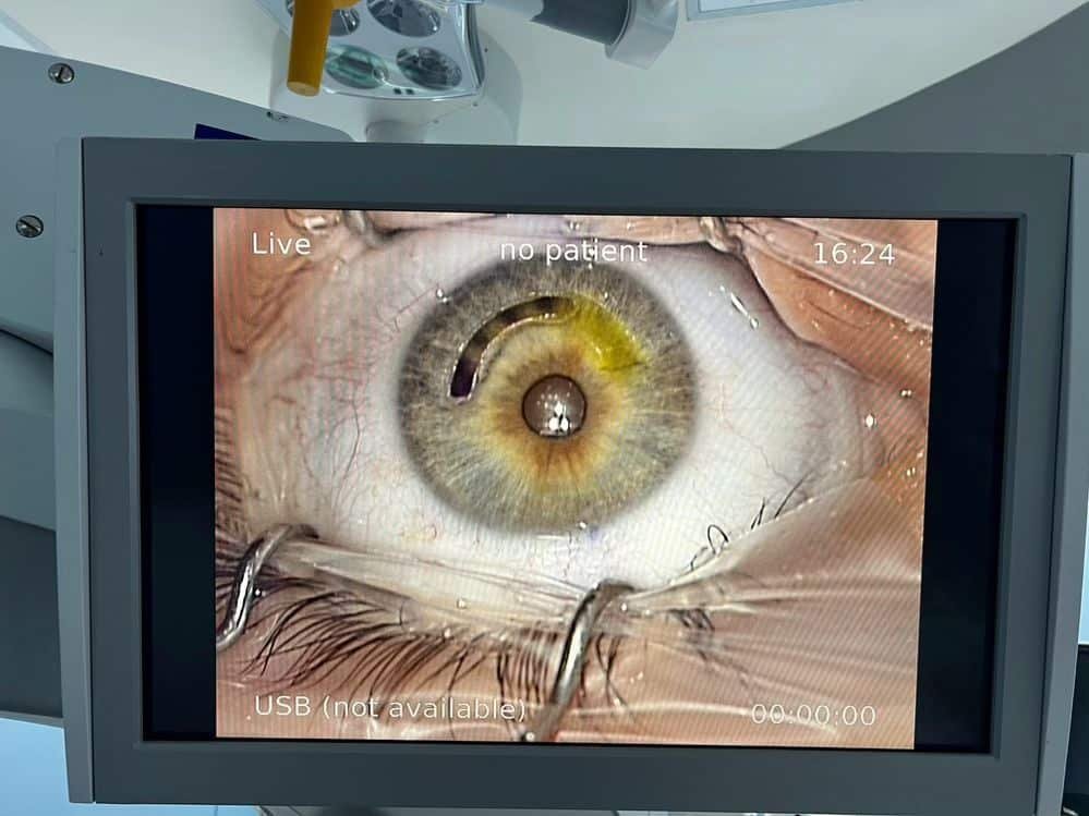
Years ago, keratoconus treatment followed a limited sequence: glasses, rigid contact lenses, and, if the disease progressed, corneal transplant. Today the situation is different. There are multiple options that can be combined depending on the patient: Cross-Linking, scleral lenses, topography-guided PRK in selected cases, intracorneal segments, Femto-CAIRS, deep anterior lamellar keratoplasty (DALK), and, as a last resort, penetrating keratoplasty.
This stepwise approach allows preserving the natural cornea for many more years and offering better visual outcomes than a decade ago. A broader review of this evolution can be read in the article on advances in keratoconus treatment.
What is Femto-CAIRS and how does it work
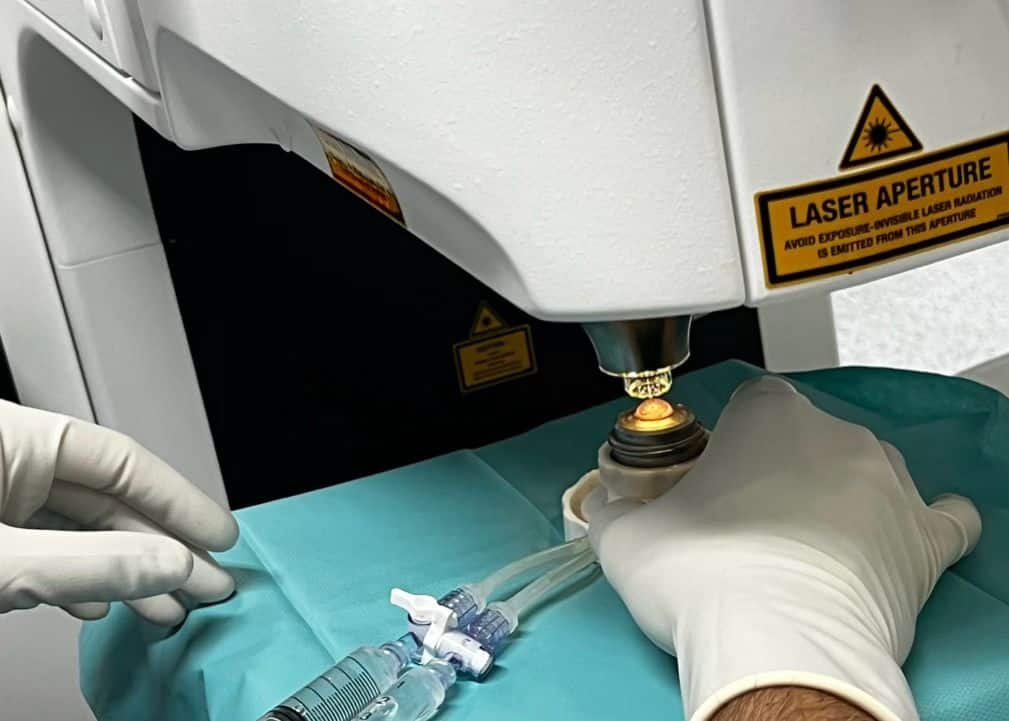
Femto-CAIRS combines two advances: the precision of the femtosecond laser to create an intrastromal channel with micron-controlled depth and geometry, and the use of segments of donated human corneal tissue instead of synthetic implants.
This second part is what makes the real difference. Traditional intracorneal rings remain as a foreign body inside the cornea. Allogenic segments, on the other hand, integrate much more naturally with the recipient tissue. Several studies published in the ophthalmic literature describe this technique: the first case series that combined CAIRS with Corneal Cross-Linking showed significant improvements in uncorrected and corrected visual acuity in patients with keratoconus stages 1 to 4.
From a biomechanical perspective, this promotes a more physiological distribution of forces within the cornea and may reduce some of the complications described with synthetic implants, such as extrusion or migration of the segment.
How Femto-CAIRS surgery is performed step by step
Femto-CAIRS is a personalized procedure: each step is adapted to the patient's corneal anatomy, and precision begins long before the operating room, with a thorough preoperative study.
After confirming that the patient is a candidate, surgery is planned using corneal topography and tomography, pachymetry, anterior segment OCT, and biomechanical analysis. These tests allow for precise knowledge of the corneal shape, tissue thickness, and ectasia location in order to design a customized treatment.
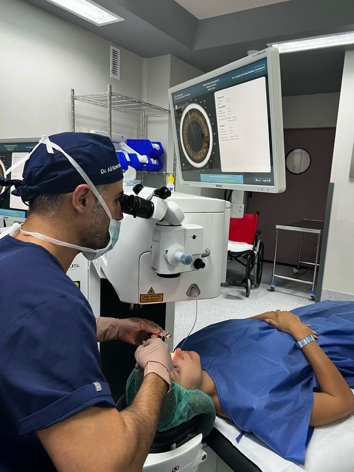
During the procedure, anesthetic drops are applied, so general anesthesia is not required and it is usually well tolerated. A femtosecond laser then creates an intrastromal channel with micron-level precision, exactly reproducing the planned depth and geometry, reducing the risk of complications associated with manual dissection.
Next, segments of donor human corneal tissue, prepared to adapt to the curvature of each cornea, are implanted. These segments reshape the cornea from within, improving its regularity and the optical quality of the eye. In many cases, Femto-CAIRS is combined with corneal Cross-Linking: while the segments improve the corneal shape, Cross-Linking strengthens the collagen fibers with riboflavin and ultraviolet light and helps stabilize the disease. The procedure lasts between 20 and 40 minutes per eye, depending on the complexity of the case.
Advantages of Femto-CAIRS compared to traditional intracorneal rings
Synthetic segments have proven effective for improving corneal shape, but as artificial implants they can be associated with extrusion, migration, infection, or intolerance. Femto-CAIRS represents a conceptual shift by using human corneal tissue instead of synthetic material. A systematic review with meta-analysis published in 2026 analyzed 18 studies with 567 eyes of 459 patients and found significant and sustained improvements in visual acuity and maximum keratometry at one-year follow-up, regardless of the segment preparation technique.
Potential advantages of this technique include:
- More natural biological integration
- Better preservation of corneal biomechanics
- Lower risk of implant extrusion
- More physiological corneal remodeling
- Better distribution of biomechanical forces
- More regular corneal surface
- Reduction of irregular astigmatism and improvement of visual quality
Although the indication must be individualized in each case, Femto-CAIRS notably expands therapeutic options for patients with moderate and advanced keratoconus, including cases with asymmetric or decentered corneas. A recent series of Customized CAIRS for decentered cones describes segments tailored according to each patient's topography, with favorable results in 32 eyes after one year of follow-up.
Femto-CAIRS and Cross-Linking: the most common combination in progressive keratoconus
Corneal Cross-Linking strengthens collagen fibers using riboflavin and ultraviolet light, and halts keratoconus progression. It is, according to a recent review on Cross-Linking techniques, the most widely used surgical procedure to stop corneal ectasia since its FDA approval in 2016.
The combination of Cross-Linking and Femto-CAIRS is especially common in patients with progressive disease: while allogeneic segments reshape the cornea, Cross-Linking stabilizes the underlying disease and reduces the risk of further keratoconus progression.
Personalized planning with Dr. Ali Nowrouzi, FEBOS-CR
No two corneas are alike, and that is one of the pillars of modern keratoconus treatment. The decision to implant allogeneic segments, perform Cross-Linking, or combine both techniques depends on multiple clinical factors specific to each patient.
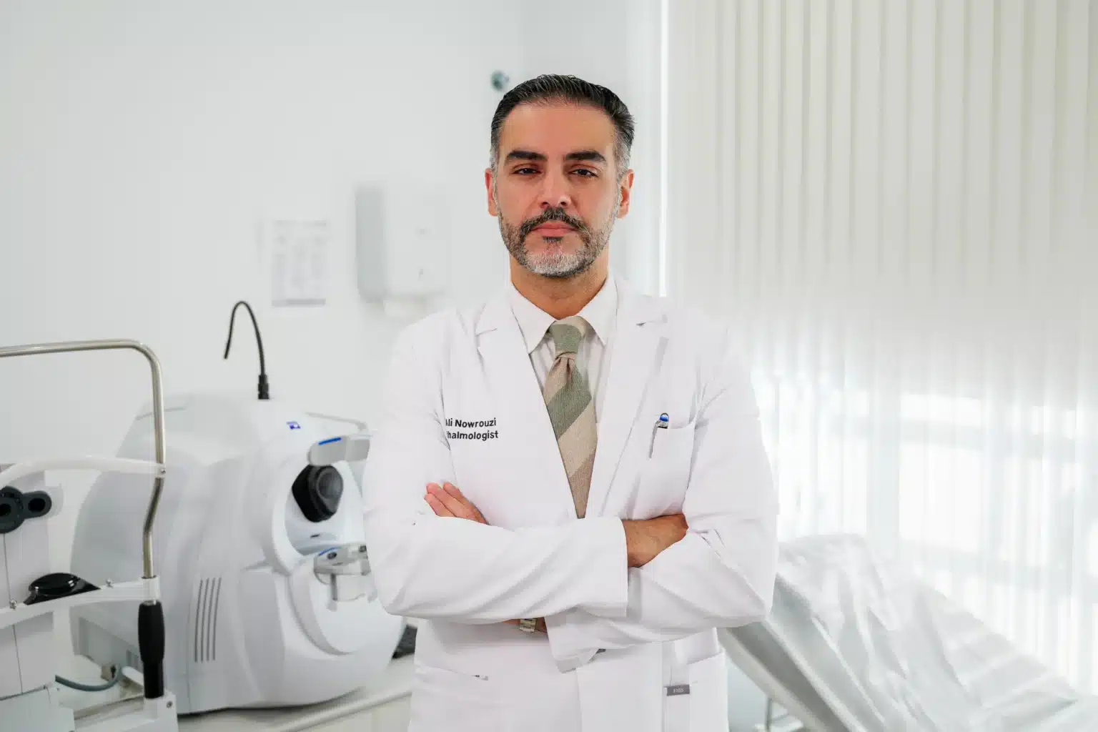
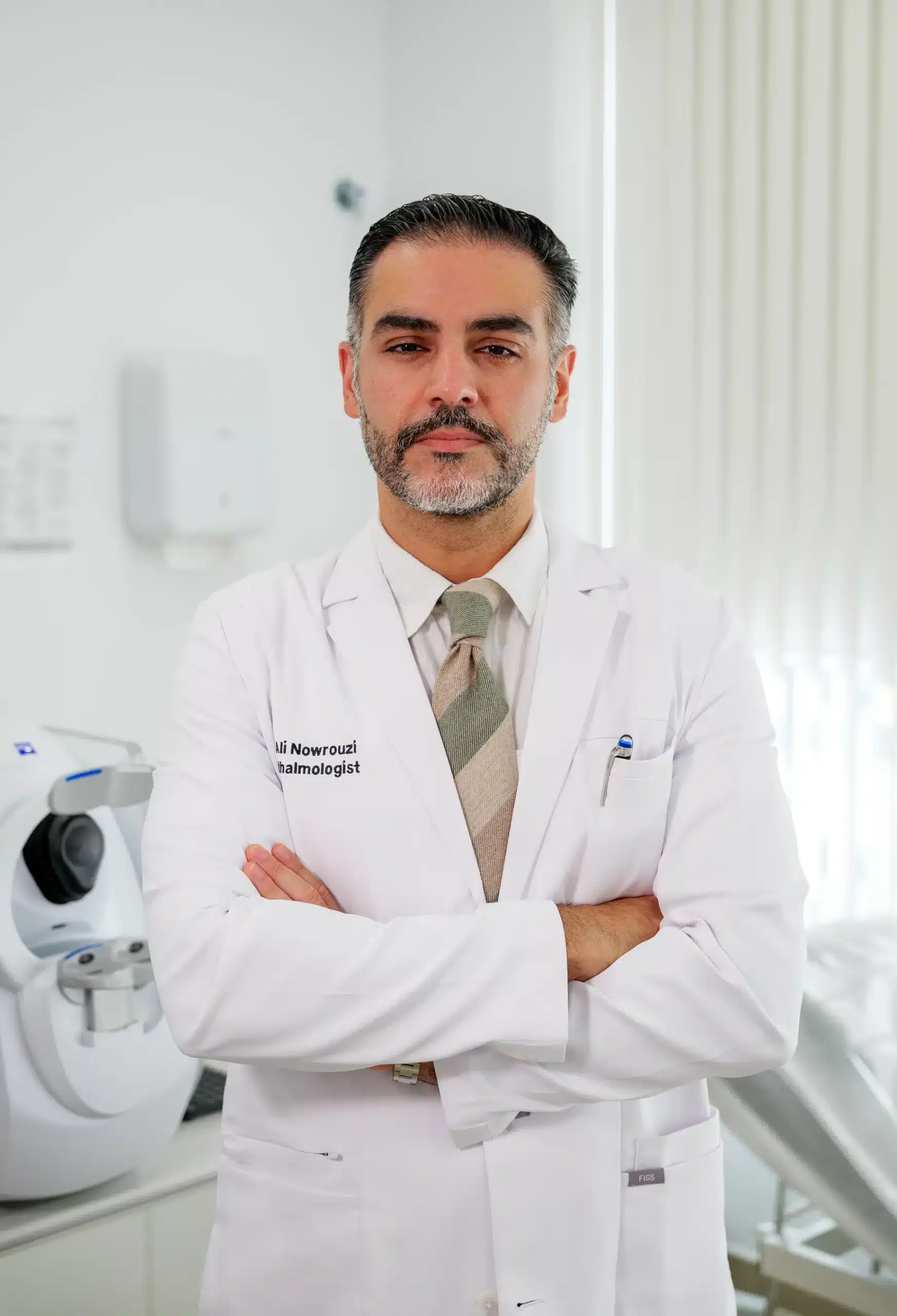
In the practice of Dr. Ali Nowrouzi, MD, FEBOS-CR, FWCRS, FACS, each case is evaluated with an advanced diagnostic protocol that studies the shape, thickness, and biomechanical stability of the cornea before proposing any treatment. The FEBOS-CR accreditation (Fellow of the European Board of Ophthalmology · Cornea & Refractive Surgery) recognizes advanced specific training in corneal and refractive surgery, complemented by the FWCRS and FACS distinctions. His complete career can be consulted in the section about Dr. Ali Nowrouzi.
This individualized planning seeks to preserve the patient's own corneal tissue as much as possible while improving their visual quality, rather than applying a standard protocol to all cases equally.
What results can be expected after Femto-CAIRS
The goal of Femto-CAIRS is not only to improve the prescription, but to restore a more regular cornea and better overall visual quality. Expected benefits include:
- Decrease in irregular astigmatism
- Improvement in corrected visual acuity
- Reduction of optical aberrations
- Greater corneal stability
- Better tolerance of contact lenses when they are still needed
- Lower dependence on more invasive treatments in selected patients
Results vary depending on the degree of keratoconus, corneal thickness, presence of scars, and other individual factors, so a specialized evaluation is essential to set realistic expectations. In patients with highly asymmetric corneas, personalized refractive surgery guided by aberrometry can complement the preoperative study.
Can Femto-CAIRS avoid a corneal transplant?
It is one of the most frequent questions in consultation. In some patients, Femto-CAIRS significantly improves corneal shape and, combined with Cross-Linking, helps stabilize the disease. This can delay or even avoid the need for a corneal transplant in certain cases.
When there is extensive corneal scarring, extreme thinning, or very advanced visual loss, a transplant may still be the best option. Each case must be assessed individually after a complete study, something that equally applies to refractive surgery in general: no two corneas respond the same to the same treatment.
Frequently asked questions about Femto-CAIRS
Does Femto-CAIRS surgery hurt? No. The procedure is performed with topical anesthesia using drops. During the procedure, the patient may feel pressure or manipulation, but typically no pain.
How long does the intervention last? Usually between 20 and 40 minutes per eye, depending on corneal anatomy and whether it is combined with Cross-Linking.
When does vision improve after surgery? Improvement is usually progressive. During the first weeks, vision may fluctuate due to the healing process, and complete stabilization may take several months.
Will I still need to use glasses or contact lenses? It depends on each case. Many patients continue to use glasses or contact lenses, although with a lower prescription and superior visual quality than before.
What tests do I need before surgery? Usually corneal topography, Pentacam, anterior segment OCT, pachymetry, aberrometry, retinal examination, complete refraction, and ocular surface evaluation.
Does Femto-CAIRS cure keratoconus? No. Currently there is no definitive cure. The goal is to improve the shape of the cornea and optimize vision, and when combined with Cross-Linking, it also aims to stabilize the disease.
Can both eyes be operated on? Yes. The strategy depends on the degree of involvement of each eye and the specialist's criteria after evaluating both separately.
Request an appointment to assess your keratoconus case in Marbella
The treatment of keratoconus requires specific experience in corneal surgery, advanced diagnosis, and personalized planning. Dr. Ali Nowrouzi is dedicated to corneal surgery, refractive surgery, and lens surgery, with a special interest in the diagnosis and individualized treatment of keratoconus at each of its stages.
If you experience frequent prescription changes, progressive blurred vision, irregular astigmatism, night halos, or a family history of keratoconus, the earlier the disease is diagnosed, the greater the options for preserving good visual quality. You can read about other patients' experiences in the section testimonies o make an appointment directly for an evaluation with advanced diagnostic tests and a treatment plan tailored to your cornea.